
GEM KNOWLEDGE · LABORATORY NOTES
Devil Aquamarine

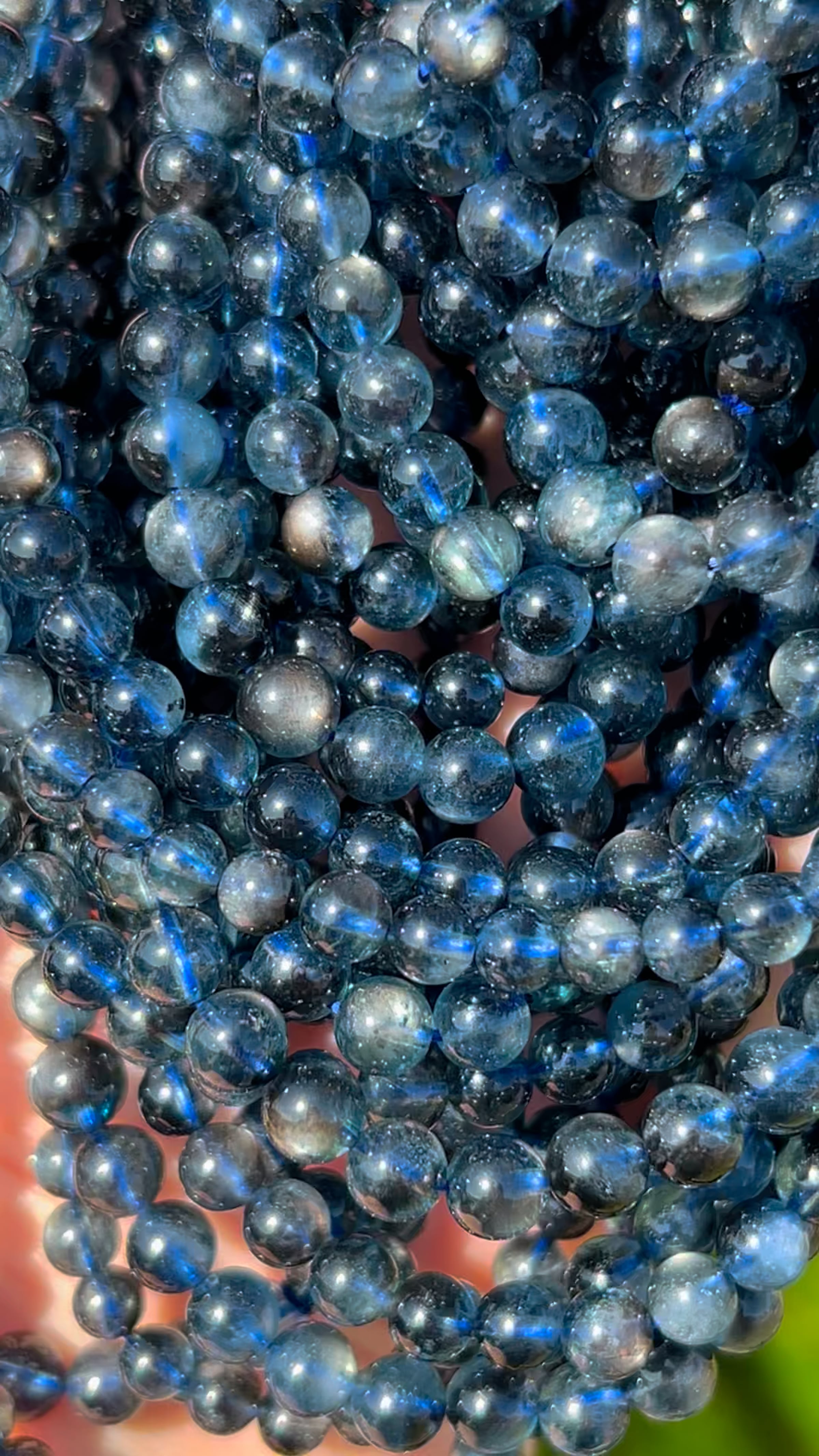

Devil Aquamarine is the supplied trade name for this blue beryl material with a striking shimmer. Soul Body Gems describes it as natural material from Brazil.
Locality and find history: Brazil. This is Soul Body Gems sourcing information.
What our testing found
X-ray diffraction confirmed beryl. Iron accounts for the blue color, and elevated sodium suggests an evolved pegmatite source. The shimmer was attributed tentatively to optical effects along healed fracture planes, rather than moonstone; no chemical difference was observed. Tiny dark mica inclusions were considered likely biotite. Some surface specks were carbon-rich, possibly epoxy.
Sample 1103, completed November 1, 2024. Findings describe the tested specimen.
View the original test results
Select a page to enlarge it.


These results are the property of Soul Body Gems, Alexander Falster, and the Maine Mineral & Gem Museum. Any mention requires full acknowledgment.
Understanding the report figures
Use this guide alongside the written findings and the original report transcript. It explains the purpose of each analytical figure and preserves the laboratory’s own figure labels. The interpretations apply to the tested specimen; signals alone do not establish the identity of every similar-looking stone.
-
Electron microscope image: This view shows small-scale regions selected for analysis. Its brightness contrast helps distinguish regions with different compositions; it is not a photograph of their natural colors. Numbered spots connect this view to the analyses listed in the report.
Laboratory figure caption: Fig.2: Backscattered electron image of an area of the specimen.
-
Elemental spectrum: The peaks record characteristic X-ray signals from elements in the analyzed spot or area. This is an analysis of that location, not automatically of the entire specimen. Any mineral identification or uncertainty named in the laboratory caption is retained below.
Laboratory figure caption: Fig. 3: Energy dispersive spectrum of spot 1 in Fig. 2. (beryl)
-
Elemental spectrum: The peaks record characteristic X-ray signals from elements in the analyzed spot or area. This is an analysis of that location, not automatically of the entire specimen. Any mineral identification or uncertainty named in the laboratory caption is retained below.
Laboratory figure caption: Fig. 4: Energy dispersive spectrum of spot 2 in Fig. 2. (carbon-rich contaminant.)
-
Diffraction scan: This scan helps identify crystalline minerals by comparing the measured pattern with reference patterns. The laboratory interpretation explains which minerals the comparison supports.
Laboratory figure caption: Fig. 5: X-ray diffraction scan of the material with beryl stick pattrn ooverlay showing match.
Read the complete report as text
Analytical Result Report
Sample id: 1103 (blue bead bracelet #5)
Submitted by: Soul Body Gems
Date completed: 11-1-2024
Operating conditions: An AMRAY 1830 SEM operated at 25 kV acceleration potential, 24 mm working distance, 0 degrees sample tilt, coated with 250 Å carbon, and also using the attached XRF unit operated at 30 kV.
A Bruker D2 Phaser X-ray diffractometer was also used and operated under 30kV and 10 mA.
Fig. 1: Photograph of the specimen. Approximately 7 mm maximum dimension of the beads. A close-up of the schiller efect is shown on the right.
Fig.2: Backscattered electron image of an area of the specimen.
Fig. 3: Energy dispersive spectrum of spot 1 in Fig. 2. (beryl)
Fig. 4: Energy dispersive spectrum of spot 2 in Fig. 2. (carbon-rich contaminant.)
Fig. 5: X-ray diffraction scan of the material with beryl stick pattrn ooverlay showing match.
Summary of findings: This specimen (Fig. 1) is pretty. The beads are made from beryl (Figs. 2, 3 and 5). The composition shows elevated sodium in the beryl which is indicative of a geochemically evolved pegmatite. The identity of beryl was confirmed with X-ray diffraction (Fig. 5). Some specks on the surface oof the beads are carbon-rich material, possibly epoxy (Figs. 2 and 4). The blue color is due to iron content (Fig. 3). The schilller effect seen (Fig. 1 right image) is probably due to optical effects on healed fracture planes. No chemical difference was noted. Some of the beads show some tiny inclusions oof a dark mica, likely biotite.
Date: 11-1-2024 Signature
Read the original laboratory report as text
The following text and tables are transcribed from the original laboratory document. Figure captions identify the report images; the laboratory interpretations and limitations are preserved.
Laboratory report 1
Analytical Result Report
Sample id: 1103 (blue bead bracelet #5)
Submitted by: Soul Body Gems
Date completed: 11-1-2024
Operating conditions: An AMRAY 1830 SEM operated at 25 kV acceleration potential, 24 mm working distance, 0 degrees sample tilt, coated with 250 Å carbon, and also using the attached XRF unit operated at 30 kV.
A Bruker D2 Phaser X-ray diffractometer was also used and operated under 30kV and 10 mA.
Fig. 1: Photograph of the specimen. Approximately 7 mm maximum dimension of the beads. A close-up of the schiller efect is shown on the right.
Fig.2: Backscattered electron image of an area of the specimen.
Fig. 3: Energy dispersive spectrum of spot 1 in Fig. 2. (beryl)
Fig. 4: Energy dispersive spectrum of spot 2 in Fig. 2. (carbon-rich contaminant.)
Fig. 5: X-ray diffraction scan of the material with beryl stick pattrn ooverlay showing match.
Summary of findings: This specimen (Fig. 1) is pretty. The beads are made from beryl (Figs. 2, 3 and 5). The composition shows elevated sodium in the beryl which is indicative of a geochemically evolved pegmatite. The identity of beryl was confirmed with X-ray diffraction (Fig. 5). Some specks on the surface oof the beads are carbon-rich material, possibly epoxy (Figs. 2 and 4). The blue color is due to iron content (Fig. 3). The schilller effect seen (Fig. 1 right image) is probably due to optical effects on healed fracture planes. No chemical difference was noted. Some of the beads show some tiny inclusions oof a dark mica, likely biotite.
Date: 11-1-2024 Signature